At CI Optometrists we are meticulous in our approach to eye care. Equipped with the latest technology great care is taken in providing a thorough check on the health and optical status of your eyes. At least 45 minutes is allocated for every eye test, allowing the optometrist time to explain any findings and address any patient concerns.
It is important to us that every client is treated as an individual and continuity of care is assured as part of the high quality service provided by the practice. We also have infection control measures across the practice for the reassurance of and safety of our clients.
If an optical correction is necessary, we will discuss your individual visual needs to ensure that the prescription we provide satisfies your occupational and lifestyle requirements.
CI Optometrists continually invest in the latest technology to provide the best quality eye care in comfortable, well appointed consulting rooms.
Digital retinal photography is undertaken as a matter of routine to assist in the detection and monitoring of retinal conditions.
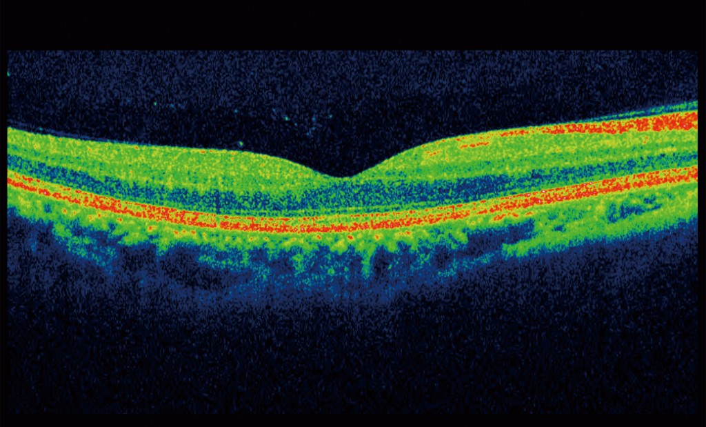
Optical Coherence Tomography – 3D OCT is a highly advanced hospital grade screening instrument which makes it possible to see beneath the surface and detect structural changes in the retina. The use of OCT has revolutionised the practice of ophthalmology in recent years, providing in vivo optical biopsy in a matter of seconds. Completely painless, non-invasive, simple and quick, it uses low coherence infrared light to take cross sectional scans, providing high resolution images of the retinal microstructure.
This has led to dramatic improvements in the understanding, early diagnosis and management of certain ocular conditions, which in turn can reduce their impact on the eyesight.
Age-related macular degeneration, diabetic retinopathy, macular holes and vitreous detachments are examples of conditions that can be detected with the OCT.
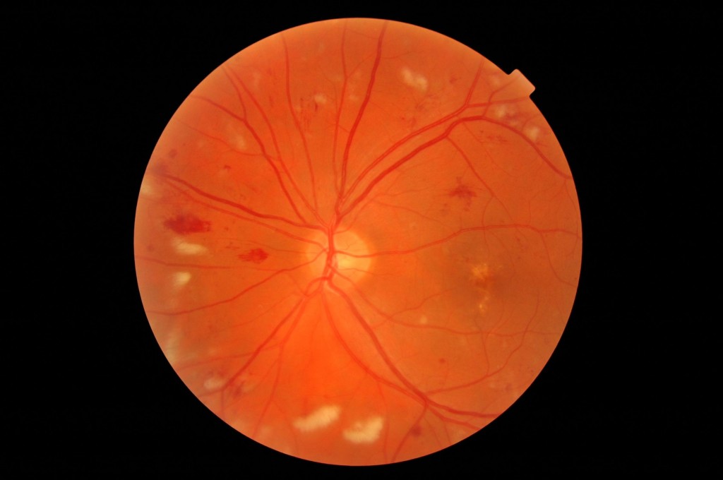
We are now offering the latest innovation in retinal technology, ultra-wide digital retinal imaging by Optos.
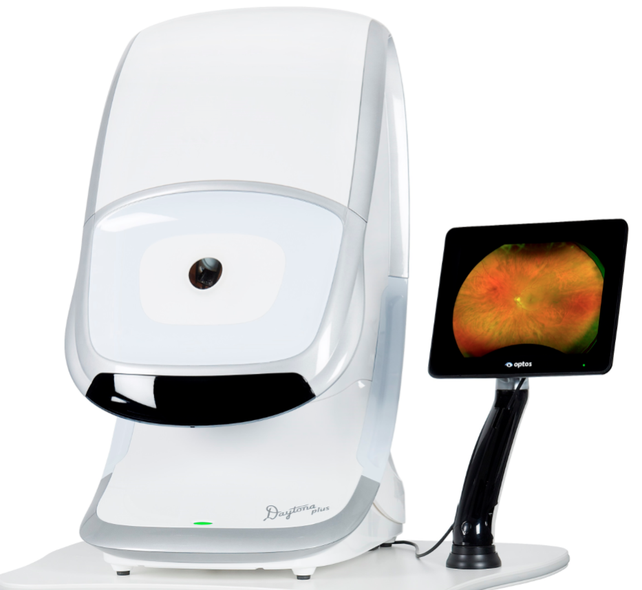
Optos Daytona Plus is a high-tech scanning laser ophthalmoscope which is used to view the peripheral retina. Optomap provides a high-resolution 200° image which is much wider than the 45° image from standard retinal cameras.
In under a second, 80% of the retina is scanned through a series of combined red and green lasers which are projected through an optical scanning system. These lasers sweep through a virtual scanning point inside the eye and then the laser light returns through the system and splits into the original red/green components. After the test, your results are immediately available for review on the screen.
Useful applications include early detection of eye problems such as retinal tears, holes or detachments. We can also detect peripheral diabetic retinal changes more easily without needing to dilate the pupil.